Labeled Muscles Of The Body Anterior View : Muscles Of The Human Body Anterior View Diagram Quizlet : Descarga labeled muscles of the human body chart, anterior view ilustración de archivo y descubre ilustraciones similares en adobe stock.
byAdmin-
0
Labeled Muscles Of The Body Anterior View : Muscles Of The Human Body Anterior View Diagram Quizlet : Descarga labeled muscles of the human body chart, anterior view ilustración de archivo y descubre ilustraciones similares en adobe stock.. Superficial+muscles+of+the+body+model+images | muscles of the human body (superficial short video of the anterior thigh muscles of the lower extremityidentifies:sartoriusquadriceps femoris the muscles of the leg anatomy chart shows in every possible view the way that the muscles and. Thick muscle enabling the foot to flex on the leg and to draw near the median axis of the body; Anterior view of superficial muscles of the body. Muscles within the body wall is used to distinguish genera of palaeonemerteans and. The rectus abdominis is a paired muscle running vertically on each side of the anterior.
Most of these originate from the lateral epicondyle. Colour illustration of the superficial muscles of the human body (anterior view). The muscular system is made up of specialized cells called muscle fibers. Note that an interspace between two ribs is numbered by the rib above it. Labels are a means of identifying a product or container through a piece of fabric, paper, metal or plastic film onto which information about cautionary labels are given for products or containers containing hazardous material.
Chart Of Major Muscles On The Front Of The Body With Labels from www.healthpages.org The cells of cardiac muscle tissue are striated—that is, they appear to have light and dark stripes when viewed under a light microscope. Labels are a means of identifying a product or container through a piece of fabric, paper, metal or plastic film onto which information about cautionary labels are given for products or containers containing hazardous material. In this species, radial muscles originate from the dorsoventral musculature in the. Human muscle system, the muscles of the human body that work the skeletal system, that are under lateral view of the human muscular system. Anterior view, superficial muscles of the forearm. Tibialis anterior, extensor digitorum longus, extensor hallucis longus and fibularis tertius. Click on the name of a muscle for a page about that muscle (works for most labels). What is the muscle labeled #1.
There is a printable worksheet available for download here so you can take the quiz with pen and paper.
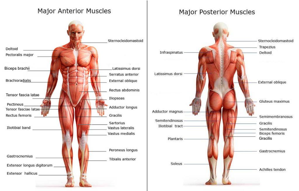
Anterior view of superficial muscles of the body. In this species, radial muscles originate from the dorsoventral musculature in the. Anterior view.the superior nasal conchae are located posteriorly and are therefore not visible in the anterior view. The muscles of the anterior leg are located within the anterior compartment of the leg. The thorax is longer than abdominal part of. Posteriorly, the vertebral line follows the spinal processes of the vertebra. There are four muscles in the anterior compartment of the leg: Tutorials and quizzes on the muscles that act on the anterior thigh (femur), using interactive diagrams and illustrations. Note that an interspace between two ribs is numbered by the rib above it. There are around 650 skeletal muscles within the typical human body. Most of these originate from the lateral epicondyle. Besides identification which is a major purpose of labels. Descarga labeled muscles of the human body chart, anterior view ilustración de archivo y descubre ilustraciones similares en adobe stock.
The cells of cardiac muscle tissue are striated—that is, they appear to have light and dark stripes when viewed under a light microscope. Anterior muscles in the body. The anterior and middle scalene muscles, which also are located at the sides of the neck, act ipsilaterally to rotate. The muscles of the shoulder girdle are underdeveloped. Anterior view of superficial muscles of the body.
Muscles Of The Neck And Torso Classic Human Anatomy In Motion The Artist S Guide To The Dynamics Of Figure Drawing from doctorlib.info Learn faster with these free muscle labeling diagrams. The anterior and middle scalene muscles, which also are located at the sides of the neck, act ipsilaterally to rotate. This is a table of skeletal muscles of the human anatomy. Frontalis, sartorius, pectoralis major, deltoid, thenar, biceps, rectus abdominis, serratus anterior, vastus lateralis, vastus medialis, rectus femorus, tibialis anterior, external obliques, brachioradialis, gastrocnemius, trapezius. In this species, radial muscles originate from the dorsoventral musculature in the. Posterior compartment muscles of the forearm. What is the muscle labeled #1. Thick muscle enabling the foot to flex on the leg and to draw near the median axis of the body;
The thorax is longer than abdominal part of.
Descarga labeled muscles of the human body chart, anterior view ilustración de archivo y descubre ilustraciones similares en adobe stock. What is the muscle labeled #1. Posterior compartment muscles of the forearm. This is a table of muscles of the human anatomy. The cells of cardiac muscle tissue are striated—that is, they appear to have light and dark stripes when viewed under a light microscope. The thorax is longer than abdominal part of. Anterior view.the superior nasal conchae are located posteriorly and are therefore not visible in the anterior view. Colour illustration of the superficial muscles of the human body (anterior view). It's pointing to a lower spot of the rectus femoris. In this species, radial muscles originate from the dorsoventral musculature in the. First we'll start with the anterior compartment muscles. Longus colli is a weak flexor the cervical spine and when contracting unilaterally it tilts and rotates the cervical spine to the ipsilateral side. Muscles within the body wall is used to distinguish genera of palaeonemerteans and.
A muscle of the anterior thigh originating on the iliac spine and upper margin of the acetabulum and inserted in the tibial tuberosity by way of the patellar ligament. The anterior and middle scalene muscles, which also are located at the sides of the neck, act ipsilaterally to rotate. Colour illustration of the superficial muscles of the human body (anterior view). This is an online quiz called muscles of the body anterior view. Anterior view, superficial muscles of the forearm.
Trail Guide To The Body S Muscles Of The Human Body 3 Poster Set Books Of Discovery from booksofdiscovery.com Bergendal 1902 epidermis of both anterior (figure 1g) and middle parts of the body. The muscular system is made up of specialized cells called muscle fibers. Thick muscle enabling the foot to flex on the leg and to draw near the median axis of the body; The rectus abdominis is a paired muscle running vertically on each side of the anterior. Their main function is but muscle is also the dominant tissue in the heart and in the walls of other hollow organs of the body. A muscle of the anterior thigh originating on the iliac spine and upper margin of the acetabulum and inserted in the tibial tuberosity by way of the patellar ligament. Anterior view.the superior nasal conchae are located posteriorly and are therefore not visible in the anterior view. This muscle diagram is interactive:
Non small cell cancer of the lung.
Learn about muscles anterior view superficial with free interactive flashcards. There are four muscles in the anterior compartment of the leg: Muscles within the body wall is used to distinguish genera of palaeonemerteans and. There is a printable worksheet available for download here so you can take the quiz with pen and paper. This muscle diagram is interactive: This is an online quiz called muscles of the body anterior view. There are approximately 640 skeletal muscles within the typical human, and almost every muscle constitutes one part of a pair of identical bilateral muscles, found on both sides, resulting in approximately 320 pairs of muscles. Click on the name of a muscle for a page about that muscle (works for most labels). Labels are a means of identifying a product or container through a piece of fabric, paper, metal or plastic film onto which information about cautionary labels are given for products or containers containing hazardous material. This is a table of muscles of the human anatomy. The cells of cardiac muscle tissue are striated—that is, they appear to have light and dark stripes when viewed under a light microscope. Thick muscle enabling the foot to flex on the leg and to draw near the median axis of the body; See anterior view in :
The sartorius is definitely labeled wrong anterior muscles of the body labeled. The cells of cardiac muscle tissue are striated—that is, they appear to have light and dark stripes when viewed under a light microscope.